Overview
Food contains carbohydrates, protein, fat, vitamins, minerals and water — none of it usable until it is broken into molecules small enough for a cell to absorb. That breakdown is the job of this whole system.
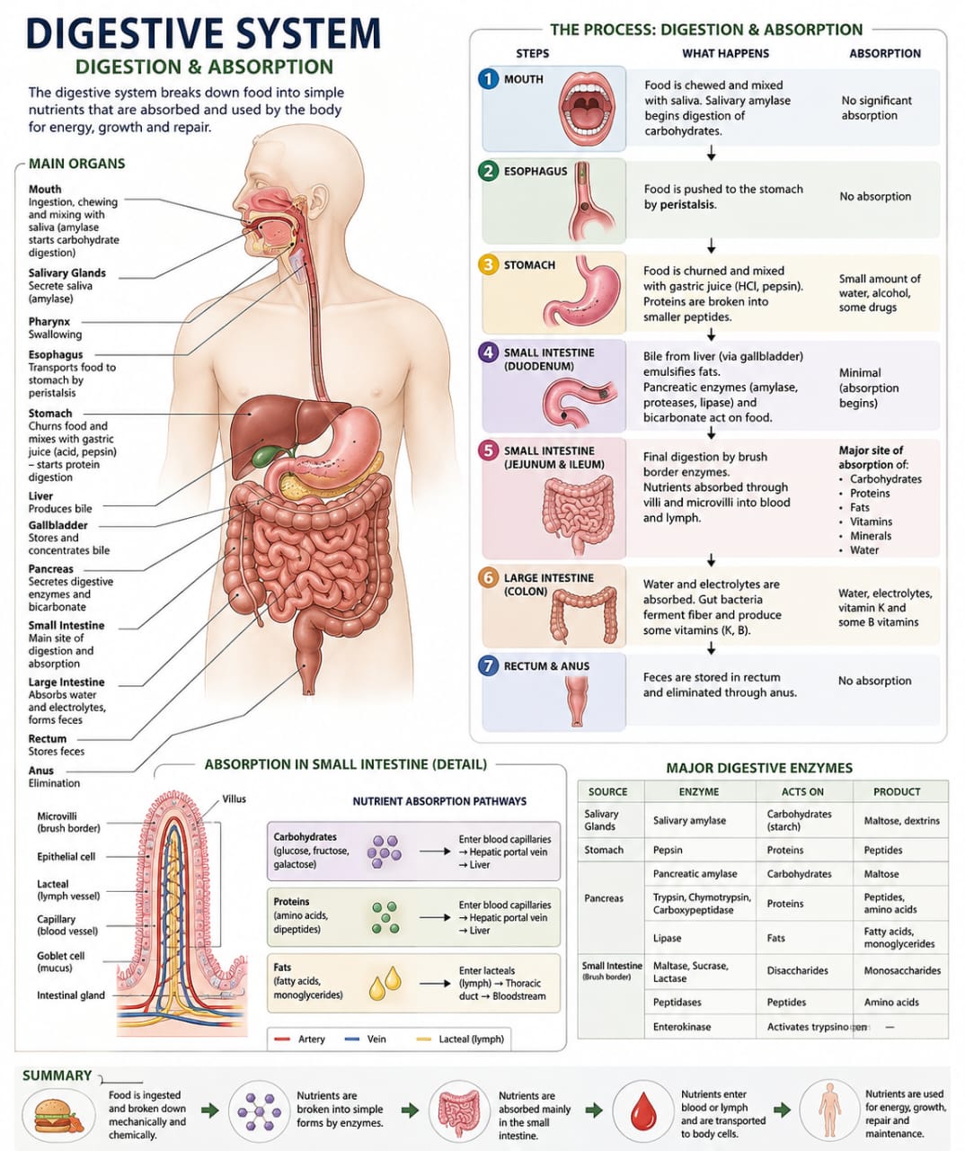
DefDigestive system
The organ system that breaks down food into simple nutrients the body's cells can absorb and use.
Two working parts — the alimentary canal (a continuous tube) and a set of accessory organs that assist but don't carry food.
Converts complex food into absorbable nutrients, then delivers them into blood and lymph for use by every cell.
Alimentary canal (GI tract)
Mouth to anus — vaayil ella bhaagam. Food physically passes through every part of this tube.
Accessory organs
Help digestion, but food never passes through them.
- Teeth
- Tongue
- Salivary glands
- Liver
- Gallbladder
- Pancreas
The five major activities of digestion
- Ingestion — putting food or water into the mouth
- Digestion — breaking complex food into simple substances
- Absorption — nutrients move from the GI tract into blood
- Assimilation — cells take up and use the absorbed nutrients
- Elimination — undigested material expelled as feces (malam)
Mouth & Oral Cavity
Digestion begins here — three things happen at once: mastication, mixing with saliva, and bolus formation.
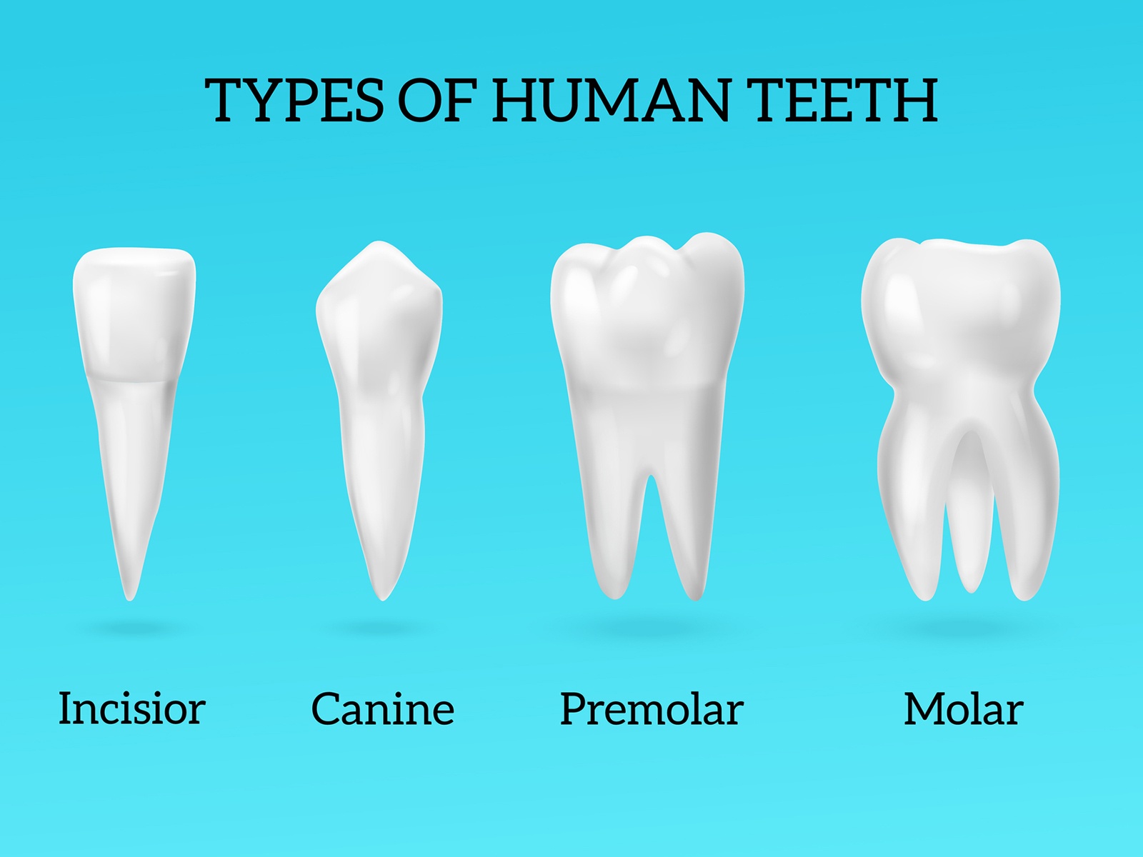
The mouth isn't just an entry point — three separate jobs happen here at once, and they overlap on purpose. Teeth cut food into smaller pieces (mastication), saliva soaks into it as you chew (mixing), and the tongue gathers the result into a soft ball called a bolus, ready to swallow.
Why bother with saliva this early, when the stomach is going to do most of the real digesting? Saliva carries an enzyme, salivary amylase, that starts breaking down starch immediately — so by the time food reaches the stomach, carbohydrate digestion already has a head start. Smaller pieces from chewing also give that enzyme more surface area to work on, the same reason chopped vegetables cook faster than whole ones.
The palate matters more than it looks like it should: the soft palate lifts during swallowing specifically to seal off the nasal cavity, which is why food doesn't usually come out your nose when you swallow. When that seal fails mid-swallow, it usually does.
The oral cavity where food enters the body and mechanical + early chemical digestion begins.
Bounded by the hard and soft palate above, teeth and tongue within, opening into the pharynx behind.
Mastication (teeth break food into small pieces), mixing with saliva, and bolus formation — the tongue collects chewed food into a soft mass and pushes it toward the pharynx.
Palate
- Hard palate — bony, rigid, anterior
- Soft palate — movable, muscular, posterior
- Uvula (annaku) — where the soft palate ends
Tongue
- Mixes food with saliva
- Pushes the bolus during swallowing
- Roles in taste and speech
Teeth
| Type | Count | Role |
|---|---|---|
| Incisor | 8 | Bite |
| Canine | 4 | Tear |
| Premolar | 8 | Grind & crush |
| Molar | 12 | Grind & crush |
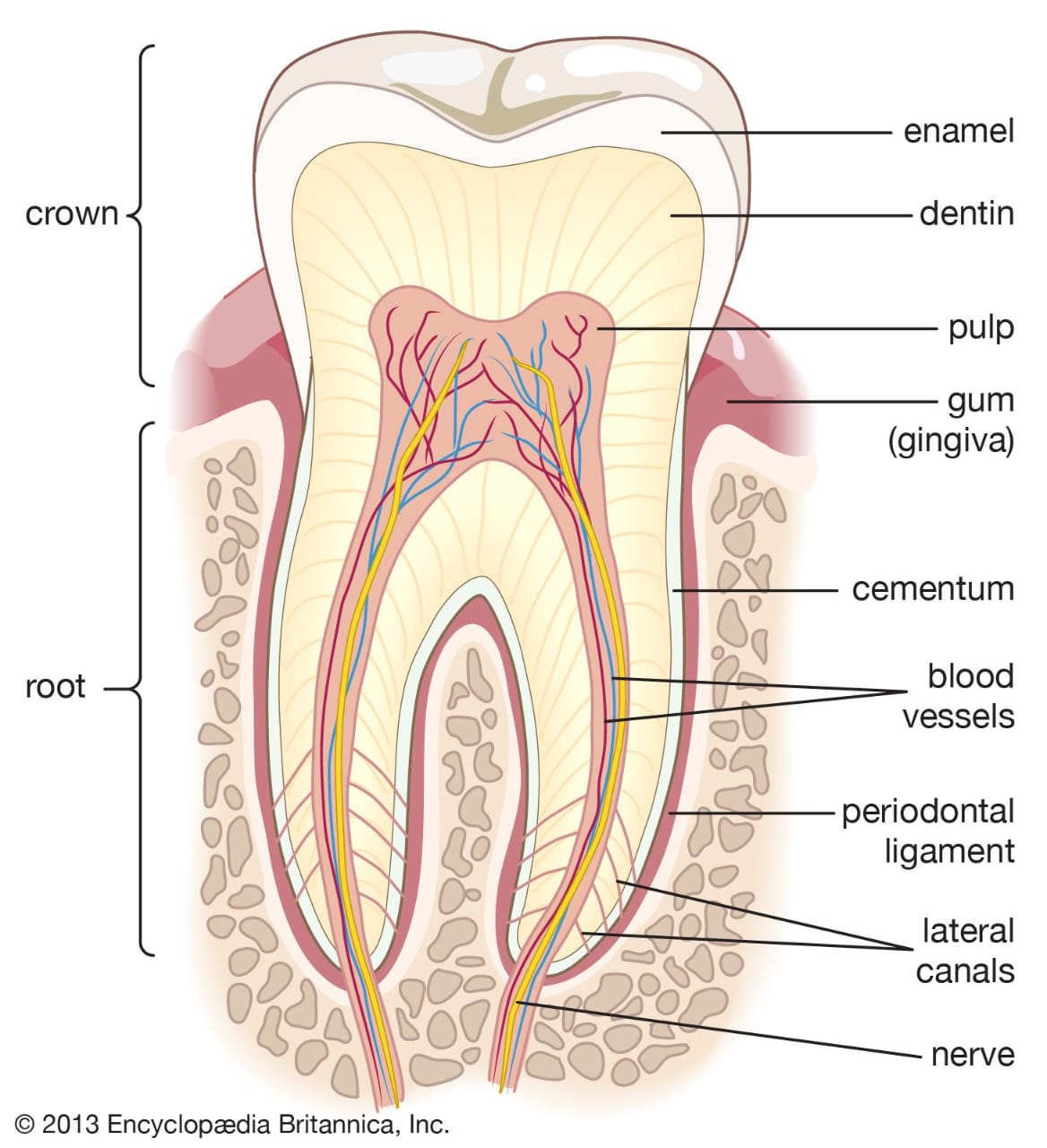
- Crown — the visible part · Root — anchored below the gum · Apex — the root tip
- Enamel — the hardest tissue in the body · Dentin — the bulk of the tooth · Pulp — vessels, connective tissue, nerves
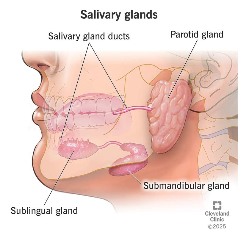
Salivary glands
- Parotid (near the ear) · Submandibular (beneath the mandible) · Sublingual (near the tongue)
Unlike humans, an elephant regrows a new set of teeth throughout life, until its final set wears out.
Your class notes don't link a named asana specifically to the mouth or salivary glands — the connection here is more general than the Section C answers for stomach or intestines.
Pharynx & Oesophagus
A shared passage, then a one-way muscular tube — this is how the bolus travels from mouth to stomach.
The pharynx is a shared corridor — the same passage carries both air and food, which sounds risky, and it is: this is the junction where choking happens if timing goes wrong. Its three regions divide the traffic: the nasopharynx handles only air, the oropharynx carries both, and the laryngopharynx is where the routes finally split, sending food into the oesophagus and air into the larynx.
Once the bolus is in the oesophagus, it doesn't just fall down to the stomach — gravity alone wouldn't work if you were lying down or upside-down. Instead, the oesophagus squeezes itself in a travelling wave called peristalsis, the same way you'd move a marble along a length of tubing by pinching and releasing behind it, one section at a time.
Pharynx: a muscular passage shared by the respiratory and digestive systems. Oesophagus: the muscular tube connecting pharynx to stomach.
Pharynx has three regions, front to back. Oesophagus is about 25 cm long.
Pharynx routes air to the larynx and food to the oesophagus without letting the two cross. Oesophagus transports food to the stomach via peristalsis — coordinated, wave-like muscular contractions.
| Region | Role |
|---|---|
| Nasopharynx | Near respiration |
| Oropharynx | Shared by food and air |
| Laryngopharynx | Leads into the oesophagus |
Stomach
A J-shaped muscular organ — part storage tank, part grinder, part acid bath.
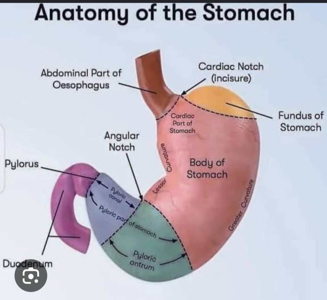
Once food is a soft, chewed mass, it drops into the stomach — and the stomach is where the body stops treating food gently. Everything here is designed to break it apart, both physically and chemically.
Physically, the stomach wall squeezes and churns like a front-loading washing machine. Its J-shape and layered muscle let it fold the food back on itself over and over, mixing it far more thoroughly than the mouth ever could.
Chemically, three different cell types in the stomach lining each add one ingredient to a deliberately harsh mix: mucous cells lay down a protective coating, chief cells release a harmless, inactive enzyme called pepsinogen, and parietal cells pump out hydrochloric acid — strong enough to dissolve metal, roughly pH 1–2.
That's also why the mucus layer matters so much: it's the only thing standing between HCl strong enough to dissolve metal and the stomach's own tissue. When that barrier weakens — from stress, irregular eating, or excess acid — you get gastritis or a peptic ulcer. This is the direct physiological reason yoga's stress-reduction practices are considered protective here: chronic stress keeps acid secretion elevated and can thin that protective layer over time.
By the time this process is done, the bolus has become chyme — a semi-fluid, acidic mix, ready to move into the small intestine in controlled amounts.
The stomach is a J-shaped muscular organ of the alimentary canal, positioned between the oesophagus and small intestine, functioning in storage, mechanical mixing, and chemical digestion.
Four regions — cardia, fundus, body (corpus), and pyloric region — with an oblique inner muscle layer and a mucosal lining containing gastric glands.
Gastric glands hold three cell types: mucous cells (protective mucus), chief cells (pepsinogen), and parietal cells (HCl and intrinsic factor). HCl activates pepsinogen into pepsin, which digests protein; the resulting acidic, semi-fluid mix is called chyme.
Also worth a line each: gastrin (a G-cell hormone that promotes gastric secretion) and intrinsic factor (parietal-cell secretion needed to absorb Vitamin B12 later in the small intestine).
Typical question: "How do asanas help in digestion?" — for the stomach specifically, the answer needs a physiological mechanism, not just a pose name.
This is the section that rewards understanding Layer 1 — you can't explain why Vajrasana or cooling pranayama help without knowing what they're counteracting.
Small Intestine
5–6 metres long, in three parts. Most digestion — and almost all absorption — happens here.
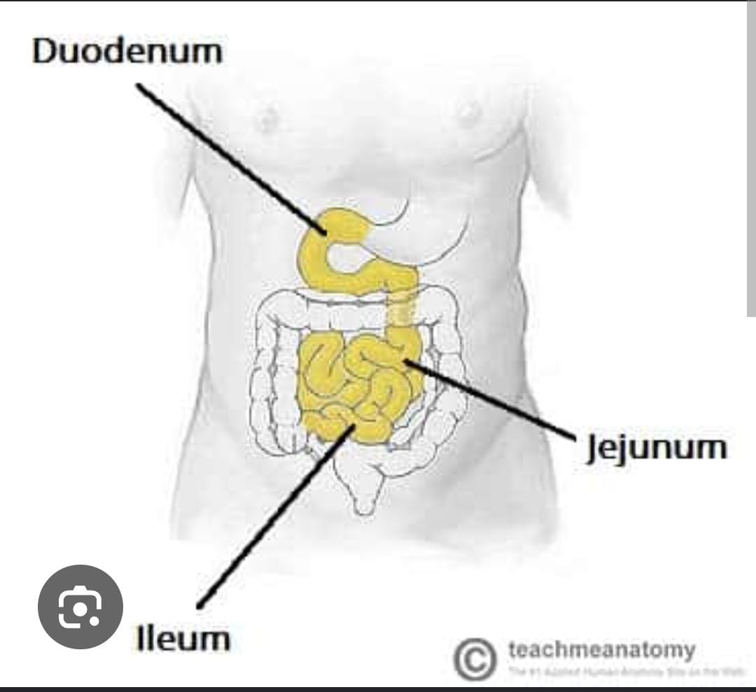
Chyme leaves the stomach in small, controlled amounts and enters the duodenum, the first and shortest part of the small intestine. This is where two other organs' outputs arrive to finish the job the stomach started: pancreatic juice (enzymes for carbs, protein, fat and nucleic acids) and bile from the liver (which handles fat). Nothing that happens later in the small intestine is possible without these two inputs meeting the chyme here first.
By the time the mix reaches the jejunum, digestion is essentially complete, and this middle section becomes the main site of absorption. The ileum, the last stretch, mostly mops up whatever's left and hands the remainder to the large intestine.
| Part | Length / position | Role |
|---|---|---|
| Duodenum | ~25 cm, connects to stomach | Receives chyme, pancreatic juice, and bile |
| Jejunum | Middle part | Major site of digestion and absorption |
| Ileum | Last part | Continues absorption, connects to the large intestine |
Villi & microvilli
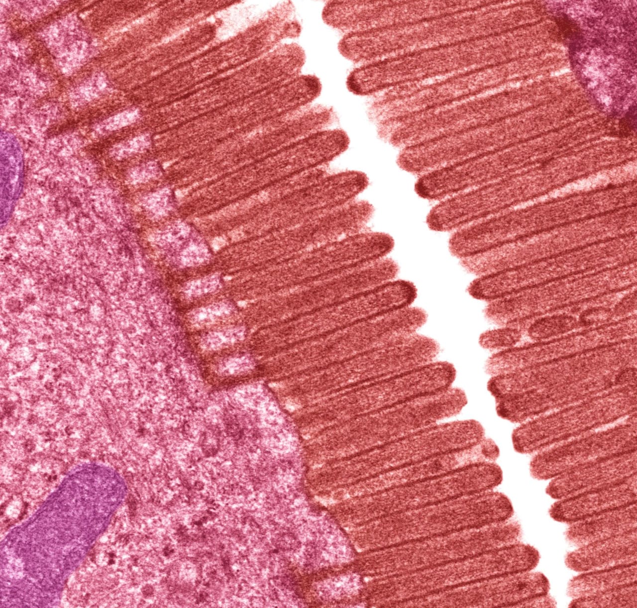
Finger-like mucosal projections lining the small intestine, each containing a blood capillary and a lacteal (lymph vessel for fat) — this is what turns the intestinal wall into a huge absorptive surface.
Pancreatic juice — enzymes by target
| Target | Enzyme | Effect |
|---|---|---|
| Carbohydrate | Pancreatic amylase | Breaks carbs toward simple sugars |
| Protein | Trypsin, chymotrypsin, carboxypeptidase | Breaks protein into amino acids |
| Fat | Pancreatic lipase | Breaks fat into fatty acids / lipids |
| Nucleic acid | Nucleases | Digests nucleic acids |
Digestion end-to-end, by nutrient
| Food | Digested by | Final product |
|---|---|---|
| Carbohydrate | Amylases + disaccharidases | Glucose (monosaccharides) |
| Protein | Pepsin + pancreatic proteases | Amino acids, small peptides |
| Fat | Bile salts + lipases | Fatty acids + monoglycerides |
Large Intestine
What's left after the small intestine is done — mostly water, electrolytes, and undigested fibre.
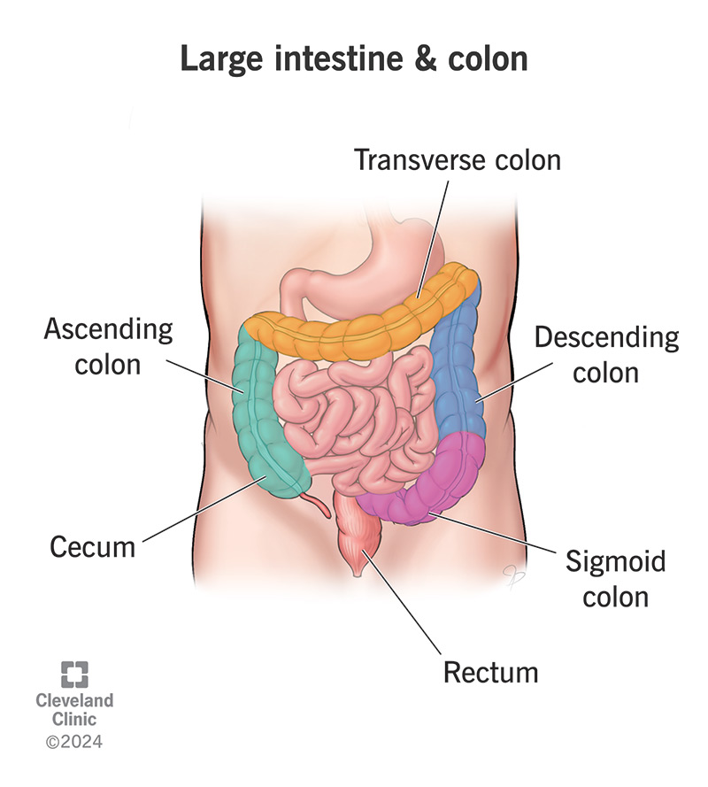
By the time material reaches the large intestine, almost every usable nutrient is already gone — what's left is mostly water, electrolytes, and fibre the body can't digest. The large intestine's job isn't digestion anymore, it's recovery and cleanup: pulling back the water and electrolytes so they aren't lost, and compacting what's left into feces.
The colon isn't sterile, either — it hosts a large population of gut microbiota, bacteria that ferment the fibre your own enzymes couldn't break down, in the process producing some vitamins (like K and certain B vitamins) that the body then absorbs as a side benefit.
Storage happens in the rectum until enough material builds up to stretch its walls — that stretch (distension) is what triggers the urge to go, technically called the defecation reflex.
- Absorption of water
- Absorption of electrolytes
- Formation and storage of feces
- Gut microbiota — bacteria metabolise undigested substances
Rectum & anus
The rectum stores feces temporarily. Distension triggers the defecation reflex — coordinated rectal contraction plus anal sphincter relaxation.
Appendix
The vermiform appendix, a narrow projection of the cecum, contains lymphoid tissue. Its inflammation is appendicitis.
Liver & Accessory Organs
The organs that support digestion without food ever passing through them.
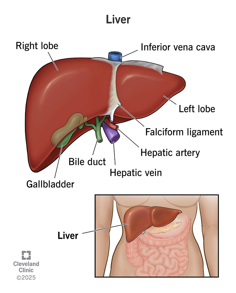
None of these three organs ever touch food directly — they're accessory organs, working from the outside by injecting their products into the duodenum exactly when needed.
The liver itself does far more than make bile — it's the largest internal gland in the body, and unusually, it can regenerate even after part of it is removed or damaged. The pancreas, meanwhile, is the source of most of the actual digestive enzymes used in the small intestine (see the Small Intestine section) as well as bicarbonate, which neutralises the acidic chyme arriving from the stomach so those enzymes can work — they don't function well in an acidic environment.
The liver is the largest internal gland in the body.
Two lobes — right and left. Its functional unit is the hepatic lobule.
Produces bile, a salty digestive juice used to emulsify fats.
Gallbladder
Stores and concentrates bile produced by the liver, releasing it into the duodenum to emulsify fats.
Pancreas
Secretes digestive enzymes (amylase, proteases, lipase) and bicarbonate into the duodenum via pancreatic juice.
Your notes don't specify a targeted asana sequence for liver disorders beyond the general obesity-linked practice above — worth asking your teacher if this comes up as a likely exam angle.
Nutrient Absorption
Each nutrient class has its own route out of the intestine and into circulation.
Once a nutrient is small enough to cross the intestinal wall, it doesn't all travel the same way. Think of it like a sorting office: water-soluble nutrients (glucose, amino acids, water itself) go straight into the blood — the fast, direct route. Fat is different: it's not water-soluble, so it can't just dissolve into blood plasma. Instead it gets packaged into fatty particles that enter the lymphatic system first (via the lacteal in each villus) and only joins the bloodstream later, further along. That's why fat absorption is structurally slower and more involved than sugar or protein absorption.
| Nutrient | Pathway |
|---|---|
| Glucose | Sodium-dependent transport into epithelial cells → blood |
| Amino acids | Carrier-mediated transport → blood |
| Water | Osmosis |
| Fat | Fatty acids / monoglycerides + bile salts → micelles → enter epithelial cells → packaged as chylomicrons → lacteals → lymph → bloodstream |
Villus cross-section — what's inside
- Epithelial cell — lines the villus, the absorbing surface
- Microvilli (brush border) — increase surface area further
- Capillary (blood vessel) — carries away sugars and amino acids
- Lacteal (lymph vessel) — carries away absorbed fat
- Goblet cell — secretes protective mucus
Common Disorders & Yoga
For each: what it is, its symptoms, and the asanas / practices the notes link to it. (Exam format: disease → symptoms → how yoga supports.)
Gastro-oesophageal Reflux (GERD) Loosened lower oesophageal sphincter
Gastritis Inflammation of the stomach lining
Peptic Ulcer Open sore in the stomach lining
Constipation Difficulty passing stools
Diarrhea Frequent, loose stools
Irritable Bowel Syndrome (IBS) Gut–brain interaction disorder
Obesity
Liver Disorders
Asanas & practices referenced across this topic
Asanas — Pavana Muktasana, Paschimottanasana, Vajrasana, Ardha Matsyendrasana, Bhujangasana, Janu Shirshasana, Pada Hastasana
Breathing / meditation — Nadi Shuddhi, yogic breathing, Bhramari, cooling pranayama, yoga nidra, cyclic meditation
Source: class notes, 10–16 Sep 2026, and Anatomy & Physiology Notes — Digestive System (PDF).